Gram Positive Bacteria Is What Color. Web gram positive bacteria are cells that take up a purple color in the gram stain procedure. Web the test, which involves a chemical dye, stains the bacterium’s cell wall purple.
If the cells take on the red colour, they are ‘gram negative’. Findings on gram stain that suggest underlying bacterial infections: Gram staining works by differentiating bacteria by the chemical and physical properties of their cell walls.
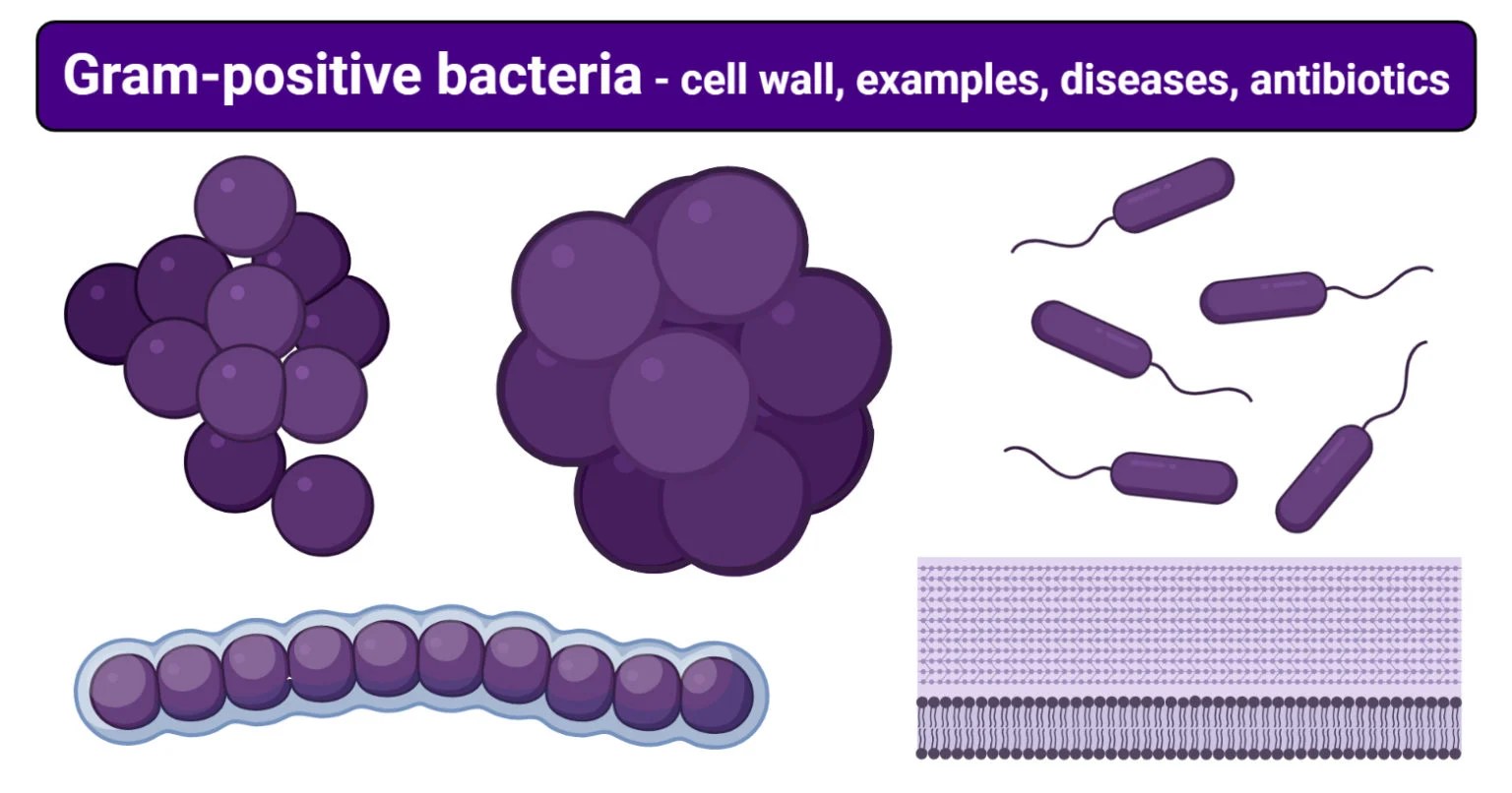
The Primary Component Of Bacterial Cell Walls Is Peptidoglycan.
Hans christian gram developed the staining method in 1884. Take a clean grease free slide. Web learn about similarities and differences in the structure of bacterial cell walls and understand why gram positive bacteria retain the purple stain while gram negative bacteria turn pink.
Web Gram Negative Bacteria Stain Red Or Pink After Gram Staining.
Web the gram stain procedure distinguishes between gram positive and gram negative groups by coloring these cells red or violet. These bacteria retain the colour of the crystal violet stain which is used during gram staining. Gram positive bacteria the cell walls of gram positive bacteria differ structurally from the cell walls of gram negative bacteria.
If The Cells Take On The Red Colour, They Are ‘Gram Negative’.
Web gram positive bacteria are cells that take up a purple color in the gram stain procedure. If the cells take on the purple colour, they are considered to be ‘gram positive’, and have a high proportion of peptidoglycan in their cell wall. Questions tips & thanks want to join the conversation?
This Difference In Coloration Is Due To Variations In.
Gram staining has been especially used because of its ability to differentiate bacteria base on their cell wall content, a major characteristic that classifies bacteria into two types. Web the organisms are identified based on color and shape. Gram positive bacteria stain violet due to the presence of a thick layer of peptidoglycan in their cell walls, which retains the crystal violet these cells are stained with.
Their Cell Wall Structure Includes A Thick Peptidoglycan Layer And Teichoic Acids.
Web the test, which involves a chemical dye, stains the bacterium’s cell wall purple. Top voted gcpuente 9 years ago around 6:00 Hans christian gram developed the staining method in 1884.